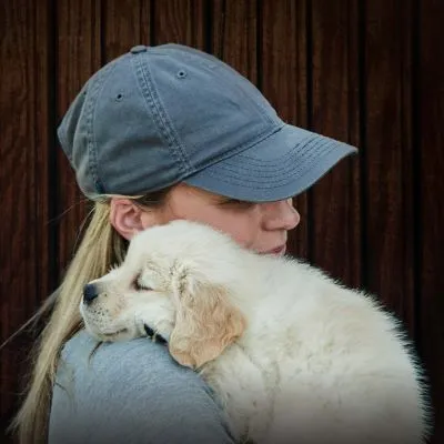
Breeder Lorraine Effa of Loriben Yorkies in Abbotsford, British Columbia, practices careful breeding by studying pedigrees and putting a great deal of thought into choosing breeding partners. Not surprisingly, she had high expectations that a particularly promising 6-month-old male, Loriben's Strike Up the Band ("Archie"), could easily become a finished conformation champion.
Effa had bred Archie's great-granddam and every subsequent generation. She knew his pedigree well. When the young dog began limping, she carefully monitored his condition. As the limping worsened, she became concerned.
After careful examination Effa's veterinarian diagnosed Archie with bilateral Legg-Calve-Perthes disease (LCPD). Radiographs showed significant deterioration of the femoral head, or the "ball" of the ball and socket, as well as muscle atrophy in both legs. Over the next year, Archie had two surgeries to ease the pain caused by the disease. Taking precaution to not perpetuate the disease in her bloodline, Effa removed Archie from her breeding program and scheduled neuter surgery for the dog.
Today, Archie is 3 years old and lives with Effa. "I have been breeding since 1995, and I've never had a dog with Legg-Calve-Perthes," she says. "It broke my heart to watch Archie go through this pain. Fortunately, he is doing very well. I wouldn't want him jumping off high places, but he can go for walks and is a sweet little pet."
After learning about LCPD research under way at Clemson University in South Carolina, Effa submitted Archie's pedigree, radiographs and a blood sample for examination. The researchers are gathering samples from dogs affected by LCPD in hopes they can accurately pinpoint a genetic marker for the disease. If a marker is found, a genetic test could possibly be developed that would lead to early detection of dogs likely to be affected and help to identify those that carry the gene mutation.
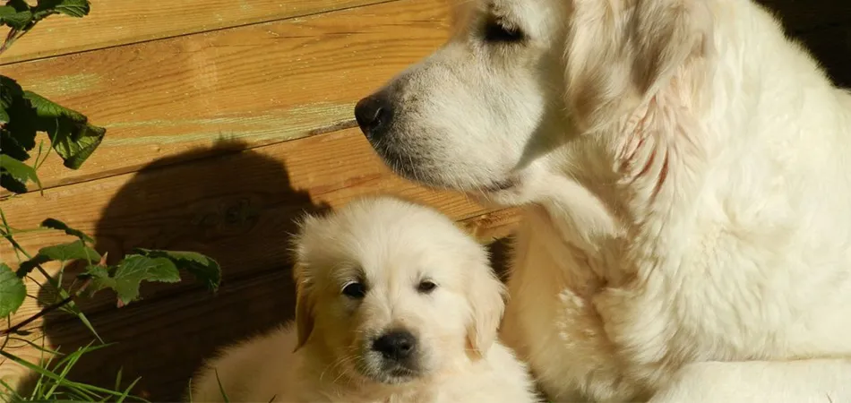
Using SNP Array Technology Yorkshire Terriers are not the only breed affected by Legg-Calve-Perthes disease. The condition, which is also known as avascular necrosis of the femoral head, affects the hip joints of several toy breeds and small terriers. According to the Orthopedic Foundation for Animals, at least 26 breeds are affected by LCPD.
Alison Starr, Ph.D., research assistant professor at Clemson University, began studying LCPD after the Westie Foundation of America contacted her for information about the disease in West Highland White Terriers. Starr, who works in the research laboratory of Keith Murphy, Ph.D., professor of genetics and chairman of the Genetics Department at Clemson, began collecting blood samples from five breeds affected by LCPD in 2007. She also obtained detailed records of each dog's pedigree and radiographs of his/her pelvis showing the hip bones.
"We wanted to study multiple breeds with the same disease presentation," Starr says. "Our goal is to determine how LCPD compares among the different breeds. Ultimately we want to learn the mode of inheritance in various breeds and be able to determine the degree to which genetics impacts disease presentation."
The research involves extracting DNA from the blood samples of affected dogs and then using SNP array technology to identify differences between the DNA of affected and healthy dogs. SNP (single nucleotide polymorphism) chips enable the researchers to pinpoint and study differences in a small area of a dog's genome rather than its entire genome.
"Hopefully the SNP chips will give us road maps to small regions on a chromosome or chromosomes to look for candidate genes," explains Starr.
"If we can identify a causative mutation or genetic marker, we can develop a test to identify at-risk dogs," Murphy says. "This would allow a breeder to make informed breeding decisions, and potentially lead to removing a particular dog from a breeding program."
In dogs with Legg-Calve-Perthes disease, an inadequate blood supply to the femoral head causes bone cells to die. The femoral head of the hip joint eventually collapses, resulting in an irregular shape and improper fit in the acetabulum, or "socket" of the ball and socket. The condition eventually leads to arthritis of the hip due to a rough, irregular joint surface.
Sudden lameness, such as the limping Effa noticed in her Yorkshire Terrier Archie, is the most common clinical sign, usually beginning between 4 and 12 months of age. Some dogs even refuse to bear weight on the affected leg; thus, over time the leg muscles may atrophy and shrink from insufficient use.
The disease may occur in both hips; however, a study by Jennifer Demko and Ron McLaughlin published in the September 2005 issue of Veterinary Clinics of North America: Small Animal Practice indicates that the disease is bilateral in only 12 to 16 percent of dogs.
Dogs suspected of having Legg-Calve-Perthes disease should have hip radiographs taken. Cara Campbell, D.V.M., who practices at the Westside Veterinary Hospital in Pearland, Texas, says veterinarians look for a flattening of the femoral head, increased space in the acetabulum or a lucency (transparency) of the femoral head.
Treatment of the disease varies according to severity of bone loss and a dog's pain. In mild cases, a dog may not experience much pain. Rest may allow healing of the damaged area. Frequently by the time a dog shows signs of pain, bone degeneration may be advanced.
"Once we see muscle atrophy in a dog, we know that pain is involved," Campbell says. "The goal is to take away the pain so they will use their leg again. If severe LCPD is left untreated, you could have bone breaks or other injuries due to a femoral head that is half deteriorated."
Surgery for Severe Cases In LCPD cases in which the femoral head is severely deteriorated, a femoral head ostectomy (FHO) is recommended. The procedure removes the femoral head and neck from the femur to alleviate pain and discomfort. A veterinary orthopedic specialist typically performs the surgery.
"The leg is put back into the approximate position it would be if there had been a femoral head," says Campbell. "Over time, the body forms fibrous scar tissue that helps to hold the bone in place."
The surgery relieves the pain caused by LCPD because it eliminates the bone-on-bone contact that can occur in the affected joint. In their research, Demko and McLaughlin found that FHO has a greater than 84 percent success rate of alleviating pain and lameness.
Campbell had FHO surgery performed on her Wire Fox Terrier, "James," to treat LCPD. "After James' surgery, we had him sleep in a 4-by-4-foot exercise pen," she says. "We restricted his activity immediately following the surgery and only allowed him to go outside to use the bathroom. By the second week, we let him resume his normal activities."
Complete recovery may take a full year. Lifetime care involves helping to avoid obesity and keeping a dog active. Though James was jumping over the baby gates at Campbell's veterinary clinic two weeks after his surgery, Campbell says it is not normal behavior following FHO surgery.
"In many cases, recovering dogs may hold the affected leg up most of the time," she says. "They may be hesitant to walk on it because the leg is going to flop around some. During recovery a dog no longer has a secure feeling of having that ball in the socket. Until the dog learns the boundaries of how steady the leg will be post surgery, he may resist using it."
Aggressive physical therapy immediately following surgery is not recommended until adequate healing occurs. Swimming and walking on a canine treadmill are safe forms of rehabilitation, Campbell says. Veterinarians also recommend manually stretching or manipulating the healing joint.
"The most important thing with rehabilitation is to be aware of any pain that may be happening," says Campbell. "You don't want to cause pain for the dog through extensive manual manipulation shortly after surgery."
Legg-Calve-Perthes disease varies significantly among dogs. Some dogs show few — if any — physical signs of the disease, while others are clearly in severe pain. Campbell was not aware that LCPD had founds its way into her breeding program until James sired two litters of puppies with the disease.
As a result of her own experience, Campbell advises breeders to have radiographs taken of every dog if LCPD is discovered in a breeding line. "Even if a dog doesn't demonstrate clinical signs of the disease, I would have radiographs taken between 6 and 7 months of age," she says. "If the X-rays look good, you should have them taken again at 10 to 12 months old. In 'Janie,' a half littermate to James, radiographic signs of LCPD didn't start showing until she was 10 months old. At that point LCPD wasn't causing pain, but still she showed radiographic signs."
Effa agrees. "As breeders, we spend thousands of dollars on training or stud fees," she says. "We can surely invest in radiographs to ensure that LCPD doesn't continue to get passed along. Contributing to Dr. Starr's research into the genetics of LCPD is also important. It's not a matter of finger pointing. It's about working together to ensure the future of our breed."
Breeds Affected by Legg-Calve-Perthes Disease
According to information on the Web site of the Orthopedic Foundation for Animals (OFA), the following toy and terrier breeds are at risk for developing Legg-Calve-Perthes disease (LCPD). The OFA maintains a database to help breeders identify dogs that test normal for LCPD. For information, visit www.offa.org.
Affenpinscher Australian Terrier Bichon Frise Border Terrier Boston Terrier Cairn Terrier Chihuahua Cocker Spaniel Dachshund Fox Terrier Jack Russell Terrier Lakeland Terrier Manchester Terrier Miniature Pinscher Miniature Schnauzer Pekingese Pomeranian Poodle Pug Schipperke Scottish Terrier Shetland Sheepdog Silky Terrier Welsh Terrier West Highland White Terrier Yorkshire Terrier
How to Participate in LCPD Research
Researchers at Clemson University in South Carolina are seeking blood samples, pedigree information and radiographs of hip bones from breeds at risk for Legg-Calve-Perthes disease (LCPD). The information will be used to identify a genetic marker for the disease. Ultimately, the researchers hope to develop a genetic test to determine normal and affected dogs as well as carriers. Information and submission forms can be found on the Yorkshire Terrier Club of America Foundation Web site at www.yorkie healthfoundation.org/samples.htm.


")