DOWNLOAD PDF
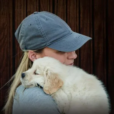
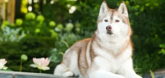
As often happens with glaucoma, “Nayla” (Cam’s Cloud In The Skeigh), a 2 ½-year-old copper-and-white female Siberian Husky, developed a severe case of the eye disease without warning. Playful and active one evening last September before bed, the beautiful, energetic dog would not get up the next morning.
“I knew immediately something was wrong,” says owner Carrie Ann Magill of Milford, Pennsylvania. “Nayla lifted her head, but she couldn’t open her right eye. It had been red for a few days, so I was concerned there was something in the eye. I thought the eye was irritated, because she’s always digging and putting her nose in chipmunk holes. When I looked into her eye, it was cloudy.”
Magill took Nayla to the veterinarian, who examined the eye for scratches and then checked the intraocular pressure. “The pressure in her right eye was over 60 compared to a normal range of 10 to 25,” Magill says. “The veterinarian told me to get Nayla to an emergency clinic right away.”
At the emergency clinic, Nayla was given oral and intravenous medications along with eye drops in an attempt to lower the pressure in the eye. Sadly, the Siberian Husky had already lost vision in her right eye.
“Her blue eye was no longer blue. It had turned brown,” says Magill. “I was in shock and broke down crying. I couldn’t believe what was happening to my ‘Nay Nay.’”
Despite the medications and eye drops, the intraocular pressure and pain continued. Five days after the glaucoma attack, Nayla had emergency surgery to remove her blind eye to alleviate the pain.
A Painful Eye Disease
Glaucoma is a group of eye diseases that occur due to abnormally high pressure in the eye. It is one of the most painful and devastating canine eye diseases, due to its prevalence, high cost of treatment and high percentage of blindness that result from the disease. Even when treated promptly, a dog often develops permanent blindness.
When a dog has glaucoma, drainage of the aqueous fluid found between the cornea and iris and between the iris and lens is impeded. The aqueous fluid helps to maintain the eye’s globular shape and nourish the eye, but when it does not drain properly, fluid builds up in the eye causing an increase in intraocular pressure. This causes pain and blocks blood flow and nerve pulses along the optic nerve. Ultimately, it causes degeneration of the retinal visual cells.
Because of Nayla’s young age, the veterinary ophthalmologist thought that possibly her glaucoma was secondary to a medical condition. Examples of causes of secondary glaucoma are: an eye tumor; luxation, or displacement of the lens; trauma; advanced cataracts; chronic retinal detachment; and inflammation of the inside of the eye causing blockage.
More often, however, glaucoma is primary or an inherited disease. The ophthalmologist examined Nayla’s eye microscopically and determined that she had primary angle-closure glaucoma (PACG), a type associated with an abnormality in the pectinate ligaments. These ligaments form a meshwork of tendrils spanning the angle formed between the iris and cornea. The tendrils normally break down into ligaments by 8 weeks of age, but when this doesn’t happen and the meshwork of tissues remains intact, the flow of intraocular fluid is hindered.
The condition is known as pectinate ligament dysplasia, or goniodysgenesis. An ophthalmologist detects the condition when performing a goniscopy test, which involves applying anesthetic drops to the cornea and using a special lens on the surface of the cornea to see inside the eye and determine the condition of the drainage angles. The test is not required for the Orthopedic Foundation for Animals’ Companion Animal Eye Registry (CAER) certification, formerly known as the Canine Eye Registration Foundation (CERF).
“It was once thought that when a dog has a normal gonioscopic examination, there is no need for further testing,” says Hans Westermeyer, DVM, DACVO, assistant professor of ophthalmology at North Carolina State University. “Now, we know that the condition can progress with age, but we are still trying to determine the relationship between the progression of these changes and the development of glaucoma.”
Importantly, most dogs with goniodysgenesis do not develop PACG; however, the vast majority of dogs with PACG have goniodysgenesis. PACG is the most common primary glaucoma in dogs and affects about 0.7 percent of dogs in North America.1 Although PACG is an inherited disease, it is genetically complex with different DNA variations attributed to causing
the eye disorder in different breeds.
Prevalence rates are difficult to establish, however, a study published in 2004 in Veterinary Ophthalmology reports much higher prevalence rates in some breeds than the average of 0.7 percent in North American dogs. Breeds with a higher occurrence are: American Cocker Spaniel, 5.52 percent; Basset Hound, 5.44 percent; Boston Terrier, 2.88 percent; Cairn Terrier, 1.82 percent; Chinese Shar-Pei, 4.40 percent; Chow Chow, 4.70 percent; Miniature Poodle, 1.68 percent; Norwegian Elkhound, 1.98 percent; Siberian Husky, 1.88 percent; and Wire Fox Terrier, 2.28 percent.
Two Genetic Studies
The AKC (American Kennel Club) Canine Health Foundation is providing funding for two separate genetic studies that aim to better understand the gene mutations that cause primary angle-closure glaucoma. In one study, researchers at the University of Wisconsin-Madison are working to identify the mutation in Siberian Huskies. At the University of California-Davis, the research focuses on finding a candidate gene for PACG in American Cocker Spaniels.
The lead investigator of the University of Wisconsin research is Gillian J. McLellan, BVMS, DACVO, DECVO, PhD, associate professor of comparative ophthalmology. She describes the work as challenging due to the seemingly complex inheritance patterns of PACG, which often involve incomplete penetrance. “Multiple genetic and environmental factors come into play in the development of glaucoma in dogs,” she says.
The two-year study, which began in March 2018, is supported by an AKC Canine Health Foundation grant of $121,740. The ultimate goal is to develop a DNA test to benefit breeders and owners.
The analysis will involve comparing the DNA of Siberian Huskies in which PACG has been confirmed after an eye has been removed due to glaucoma with Siberian Huskies over 10 years of age with no signs of glaucoma based on an ophthalmological examination and gonioscopic test.
Genotyping of DNA samples precedes a genome-wide association study using genomic technologies. “We will be leveraging technologies and approaches that provide a more powerful genetic approach than techniques used in the past. This includes using a newly developed very high-density array to detect genetic variation in canine DNA and rapid sequencing of the whole genome of selected animals,” explains Dr. McLellan. “Once a potential gene of interest is identified, we will screen the DNA of the general Siberian Husky population to confirm the gene’s association with disease.”
Identifying the genetic mutation(s) will help breeders make informed breeding decisions, which is particularly important since affected Siberian Huskies generally develop glaucoma around 5 ½ years of age, after they’ve been bred, though the breed can be affected by glaucoma early in life. A DNA test also would help identify dogs at risk of glaucoma, which is important since there seldom are signs preceding a PACG attack.
“Knowing a dog is susceptible to PACG can put owners on alert, so they can begin preventive treatments that may thwart an attack as well as help them be better prepared to deal with an attack,” says Dr. McLellan. “Usually veterinary ophthalmologists begin prophylactic treatment only after the first eye is lost to glaucoma. Knowledge of a dog’s genetic status will hopefully help prevent blindness from glaucoma in the future.”
A genetic test would potentially reduce reliance on gonioscopy testing to identify dogs with a greater risk of developing PACG. Dr. McLellan cites limitations of gonioscopy testing, which include a lack of consistent grading of severity among veterinarians and an inability to view structures deeper in the eye than the pectinate ligaments that could contribute to blockage of aqueous fluid outflow.
“Additionally, gonioscopy findings are not stable throughout a dog’s life, meaning that a dog’s glaucoma risk status can increase with age,” she says. “Notably, some dogs with a severe grade of goniodysgenesis might never develop glaucoma.”
Meanwhile, the study of PACG in American Cocker Spaniels underway at the University of California-Davis involves identifying genetic markers that cause the disease in the breed most commonly affected by glaucoma. Sara Thomasy, DVM, PhD, DACVO, associate professor of comparative ophthalmology, is leading the research.
Funded by the AKC Canine Health Foundation, the two-year, $40,000 study, which began in May 2017, involves comparing DNA samples from affected Cocker Spaniels with controls. The study includes 40 affected dogs and 85 controls. As with the glaucoma research in Siberian Huskies, the goal is to develop a genetic test to aid breeders in their breeding programs.
“We are about to submit the first samples for genetic analysis,” says Dr. Thomasy. “We have carefully phenotyped the cases and controls using gonioscopy and ultrasound biomicroscopy to view the drainage angle. We also used optical coherence tomography to assess the health of the retina and optic nerve. Should we find a candidate gene, we will need additional cases of affected American Cocker Spaniels to verify the result.”
In all dogs, glaucoma in one eye is almost always followed weeks or months later by glaucoma in the other eye. Because of this, Nayla, the Siberian Husky, was checked with gonioscopy one month after losing her right eye to PACG. The test showed that her left eye was at high risk for glaucoma.
“Nayla’s ophthalmologist began giving her prophylactic therapy to try and delay the onset of glaucoma in the eye,” Magill says. “This may help delay the onset of glaucoma for up to 30 months, but at some point, possibly sooner, the left eye will develop glaucoma. Once the pressure increases in the left eye, we will only have hours to bring it down to normal or she will lose her vision.”
In the meantime, Magill plans to send Nayla’s cheek swab with her DNA to Dr. McLellan’s study of PACG in Siberian Huskies. “Every day I am hopeful that the preventive treatment will work,” she says. “Nayla has been through so much. If we can help the researchers find the gene that causes this horrible
disease, this would mean the world to me.” n
1Gelatt KN, MacKay EO. Prevalence of the Breed-Related Glaucoma in Purebred Dogs in North America. Veterinary Ophthalmology. 2004;7(2);97-111.
Siberian Husky owners Can Contribute to Research
Genetic samples of DNA from Siberian Huskies with a history of primary angle-closure glaucoma as well as from healthy Siberians over 10 years old are needed for research at the University of Wisconsin-Madison. A cheek swab containing a dog’s DNA will help the research team as they genotype DNA samples from affected dogs and healthy control patients to complete the initial genome-wide association study. To learn more, visit this link or call 608-263-7600. To help support the study, visit this link.
Signs of Glaucoma
• Redness of the white of the eye
• Color change to the eye
• A dilated pupil that doesn’t respond to light
• A cloudy eye
• Intermittent blindness
• Squinting
• Discharge from the eye
• Behavior changes, such as being less active
You should contact your veterinarian immediately if you recognize these signs of glaucoma in your dog. To find a board-certified veterinary ophthalmologist, search the website of the American College of Veterinary Ophthalmologists
Understanding Glaucoma Treatment & Disease Progression
Time is critically important in getting a dog suspected of glaucoma to the veterinarian. Sadly, most dogs have lost their vision by the time they are examined by a veterinarian.
Treatment for dogs at risk of glaucoma may include topical and systemic anti-glaucoma medications. These preventive medications, which are prescribed on a lifelong basis, help to decrease the intraocular pressure by reducing the production of aqueous fluid or increasing its outflow. In some cases, they may help vision return.
In addition, the intraocular pressure in the eye must be regularly monitored. It helps to avoid stress and to use a harness rather than a collar when walking an affected dog.
“The mainstay of glaucoma therapy is eye drops,” says Hans Westermeyer, DVM, DACVO, assistant professor of ophthalmology at North Carolina State University College of Veterinary Medicine. “These eye drops are the same drops used to treat glaucoma in humans. The monthly cost can be around $60 to $100.”
As the disease progresses, increasing amounts of medications are needed to help keep the intraocular pressure in a healthy range. Eventually, the disease progresses to the point the only recourse is surgery.
If the eye still has vision, a veterinary ophthalmologist considers two surgeries — endolaser cyclophotocoagulation or the surgical placement of a shunt — in determining which is best for an individual dog. Endolaser cyclophotocoagulation uses a laser beam to destroy cells that produce the aqueous fluid and requires concurrent cataract surgery and artificial lens placement in the eye. Glaucoma shunt surgery involves placing a drainage device, a tiny shunt, into the eye to help drain the aqueous fluid.
Unfortunately, even these surgeries do not represent a cure. On average, they are only able to control the glaucoma for one to two years. They also are expensive, with costs in the thousands of dollars.
If a dog has already lost its vision, removal of the eye is generally recommended to reduce the pain and pressure. An intraocular prosthetic eye can be implanted, though considerations include the possibility of corneal abrasions and dry eye.
To better understand the ocular changes that occur over time in dogs with primary angle-closure glaucoma (PACG), Dr. Westermeyer has begun a study that involves identifying the anatomical features that will help to predict when a glaucoma attack is going to occur. The research is funded by the American College of Veterinary Ophthalmologists’ Vision for Animals Foundation.
“PACG in dogs is a devastating disease because the attacks of extremely high intraocular pressure occur suddenly and unpredictably,” Dr. Westermeyer says. “This study involves following dogs with confirmed PACG in one eye to determine if there are changes that will predict the onset of elevated pressures in the second eye.
“We hope to not only learn how to predict when glaucoma will develop in the second eye of a dog that already has been diagnosed with glaucoma, but also how to predict which dogs will develop glaucoma in the general dog population.”
The study, which began in December 2017, has enrolled seven dogs with PACG, with a goal of enrolling 21 dogs with glaucoma and seven controls. The research team continues to enroll dogs that have developed glaucoma within the preceding month, have a normal opposite eye, except for goniodysgenesis, and are otherwise healthy. Participating dogs will receive free bloodwork and undergo free ophthalmologic examinations every three months. For information, contact Dr. Westermeyer at hdwester@ncsu.edu
or 919-513-6659.
Cocker Spaniel Owners Can Help Advance Glaucoma Study
If you have a healthy American Cocker Spaniel that is at least 10 years of age with normal eyes or one diagnosed with primary glaucoma, you may be able to participate in a genetic study underway at the University of California-Davis. To learn more, go to this link. To help support the study, visit this link.


")